The MIRCOM microbeam
MIRCOM, the ion microbeam for radiobiology of intra- and intercellular communications, is an irradiation platform with an ion microbeam capable, with micrometric precision, of targeting cellular or subcellular elements with a defined number of charged particles. It is an essential tool for IRSN’s radiobiology research. Among other things, this research has applications in the fields of ionizing radiation use for diagnostic and therapeutic purposes, and manned spaceflight.
Background of the research
In oncology, progress with diagnosis and treatment has increased the chances of cure for many patients. However, it has not yet succeeded in eliminating the after-effects of either the disease or the treatment, for affected patients. Efforts are under way to reduce treatment toxicity through research and development of personalized medicine. IRSN is contributing to this by developing research into the biological mechanisms involved in the initiation and progression of these side-effects, so as to be able to quantify their risk of occurrence and offer new therapies for treating them. In the arsenal of available technologies that involve the use of ionizing radiation, hadron therapy (radiotherapy using proton or carbon beams) represents a promising step forward. It requires new, more specific, dosimetry concepts to be developed, tailored to the biological efficacy of the particles, which is different from that of the photons used in traditional radiotherapy. To do this, better knowledge is needed of the links between the initial events related to the transfer of energy by the ionizing radiation at cellular level, and their early and delayed biological effects.
Issue of research around MIRCOM
IRSN’s ROSIRIS program aims to use an experimental approach on the MIRCOM platform to fill part of the gap in existing knowledge between the physics of ionizing radiation and the initial events induced at molecular, cellular and tissue level. The research will establish correlations between the topology of energy deposits by the ionizing radiation (protons, alpha particles, carbon ions, etc.) on a nanometric scale and radiation-induced biological events.
Conventional irradiation tools irradiate the whole of a sample, with random statistical distribution of the ionizing particles. This makes it impossible to know whether each cell in the sample has actually been irradiated and to target a sub-compartment of the cell. However, this is possible with a microbeam, since it offers the possibility of delivering a predetermined number of particles of defined characteristics (type and energy) to a specific area of the cells, the nucleus or the cytoplasm, with a spatial resolution of around a micrometer. The microbeam’s precision therefore means that the fraction of the cell targeted by the particles, the number of particles for each target, and their location at cellular or sub-cellular level, can be known.
The MIRCOM microbeam
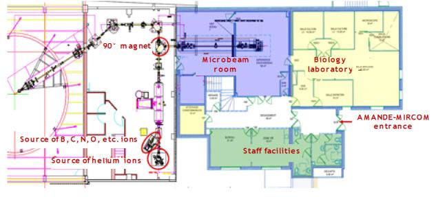
The MIRCOM microbeam, installed on IRSN’s Cadarache site (Bouches du Rhône), is ‘grafted’ onto the AMANDE facility’s accelerator, which can create monoenergetic neutron fields for use in neutron metrology and dosimetry.
Ion sources
The accelerator of the AMANDE and MIRCOM facilities has a total of three sources of negative ions delivering ion beams ranging from several tens (helium ions and heavier) to several hundreds of µA (in the case of hydrogen and deuterium):
- A ‘multi-cusp’ ion source, originally installed to meet the AMANDE facility’s needs, which can generate hydrogen and deuterium ions (Z = 1). Only the hydrogen ions are used for MIRCOM’s requirements.
- A second ‘multi-cusp’ source for producing helium ions (Z = 2).
- A third source (cesium sputter source) for producing ions above boron (Z = 5). The main ions that will be used are boron (Z = 5, carbon (Z = 6) and oxygen (Z = 8).
Operation
The ion beam is accelerated in a 2MV TandetronTM. The negative ions are accelerated to an energy of 2 MeV maximum in the center of the Tandetron, where they are ‘stripped’ to make them positive and accelerated again up to an energy dependent on their charge. The ion beams produced have a maximum energy of 4 MeV, 6 MeV or 8-10 MeV for the protons, alpha particles or B, C and O particles respectively. The beam of accelerated ions is then bent using a 90° magnet towards the microbeam line. This magnet can select the ions to be used for the irradiation based on their mass, charge and energy. They then pass into an ‘object’ box consisting of two successive stages of tungsten jaws (horizontal and vertical) and a collimator stage, where the beam size and intensity are reduced so only the core is kept.
Downstream of the object box, the beam is only a few nA, compared to several µA upstream of it. The beam then passes through a collimator to eliminate the particles scattered a long way from the beam’s optical axis and thus to limit the aberrations induced by the focusing system: the final intensity is 1000 to 15,000 particles per second, depending on the collimator used.
After this second collimation, the beam passes through a focusing system consisting of four magnetic quadrupole lenses, coupled to ensure the ion beam is focused symmetrically. This system can achieve a theoretical enlargement factor of around 1/20, i.e. the beam obtained under vacuum after focusing is approximately 20 times smaller than the object collimator used.
An electrostatic deflector, placed upstream on the line, can cut the beam in a few microseconds after a given time on the target or, coupled with an ion detection system, when the required number of particles has reached the target.
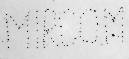
Just before its extraction in air, an electrostatic displacement system is used to precisely position the beam on the target. This system can move the ion beam from one point to another in a few microseconds, and can thus generate different irradiation patterns (single point, grid, cross, etc.).
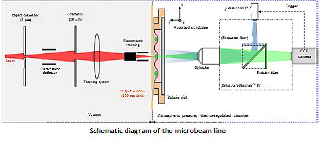
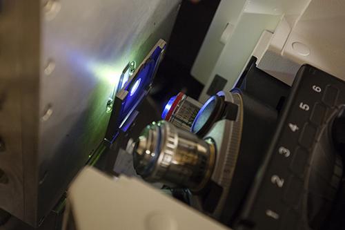
The beam leaves in the atmosphere through a 150 nm thick extraction window made from Si3N4 opposite which there is a motorized sample holder and an epifluorescence microscope. The various systems are managed by specially developed software that automates the calibration and irradiation phases. One special characteristic of this microbeam is that, because of its microscopy system, it can perform in-line video microscopy, which means that radiation-induced effects occurring in the samples can be seen directly only a few seconds after irradiation.
To overcome mechanical vibrations as far as possible, the key elements of the device (object box, focusing system, microscope) are placed on massive marble blocks mechanically isolated from the rest of the building.
Characteristics
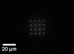
4 MeV proton beam with a diameter under vacuum estimated at less than 1 µm
Estimation of performance on target :
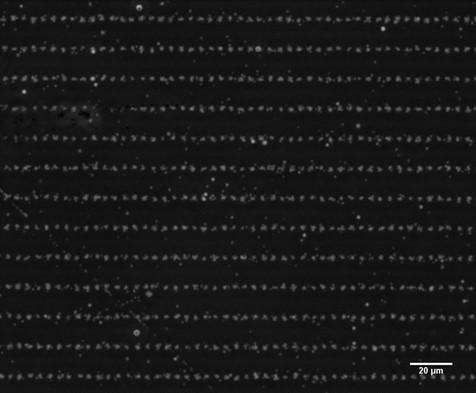
4 MeV proton beam on track detector. 10±3 ions per point. Step: 5x15 µm.
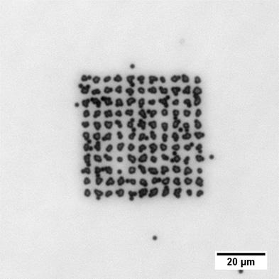
6 MeV alpha particle beam on track detector. 10±3 ions per point. Step: 5x5 µm
Beam of 6 MeV alpha particles on a trace detector - Targeting with 10±3 particles per point.

Beam of 6 MeV alpha particles on a trace detector.
Biology laboratory
MIRCOM has a 120 m² biology laboratory equipped with everything needed to culture cells and analyze the effect of radiation. It is next to the microbeam room and means that biological samples can be quickly and easily transferred to and from the microbeam. It consists of:
- 2 L2 containment rooms (with biosafety cabinet, thermostatically-controlled incubator, refrigerated centrifuge, microscope, refrigerator, etc.);
- 1 microscopy room with a Zeiss inverted fluorescence microscope with a thermostatically-controlled and gas- (CO2) and humidity-regulated enclosure for time lapses of varying lengths;
- The Definite Focus module maintains the focus regardless of the experimental and environmental conditions. The illumination sources are an X-Cite 120 LED source and filters for Dapi, GFP, TxRed and Cy5.
Objectives:
* LD Plan-Neofluar 20x/0.4 Ph and 63x/0.75
* Plan-Apochromat 10x/0.45 Ph, 20x/0.8 Ph and 63x/1.4 Oil
It has an Apotome 2 Structured Imaging Module camera and a Hamamatsu Orca Flash 4.0 V2+ camera.
MIRCOM – a platform open to the scientific community
The MIRCOM microbeam line was developed by the Center for Nuclear Research at Bordeaux-Gradignan (CENBG), a CNRS/IN2P3 and University of Bordeaux establishment, with which IRSN has developed a close working relationship. The line complements CENBG’s line at the AIFIRA platform, which has a 3.5 MV Singletron accelerator that produces proton, deuteron and alpha particle beams.
MIRCOM is open to research teams from the national and international scientific community, especially Europe, in the context of radiation protection research programs.