The SARRP irradiator
The SARRP irradiator (Small Animal Radiation Research Platform) is an image-guided microirradiation system, delivering low energy X-rays, for small animals. The IRSN uses it to conduct preclinical research on the effects of irradiation on healthy tissue and its treatment. In particular, this irradiator is dedicated more specifically to the fields of radiobiology, the adverse effects of interventional radiology, the adverse effects of high doses of radiation delivered during certain types of radiotherapy, especially stereotactic radiation therapy.
Background and objectives
One of the IRSN’s missions consists of evaluating the risks related to the use of ionizing radiation in the medical sphere and in accidental situations. The Institute therefore conducts research programs in partnership with French and foreign universities or other research bodies:
- on the potential adverse effects of interventional radiology, which consists of performing radiology image-guided or image-controlled (radiography, radioscopy) invasive medical procedures (puncture, biopsy, surgery, etc.);
- on the adverse effects of high doses delivered during stereotactic radiotherapy, which consists of treating highly localized cancer sites;
- on acute radiation syndrome.
To study the biological mechanisms of irradiated tissue, the IRSN acquired a SARRP microirradiator in 2015, a device delivering targeted irradiation for small animals (rats, mice).
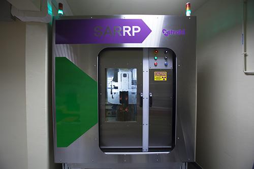
Description of the facility
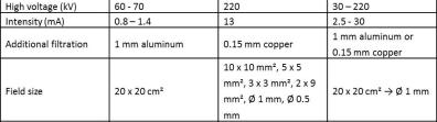
The SARRP is an image-guided microirradiator using a conventional X-ray source (X-ray tube, 30 – 225 kV), mounted on an isocentric gantry which revolves at more or less 180°and which produces both the irradiation and the image. In effect, with an imaging system inside the irradiation cabinet, it is possible:
- to capture high resolution (200 µm) scan images of small animals (rats, mice),
- to target a specific anatomical structure of the animal (accuracy of 0.2 mm) via the Muriplan treatment planning system,
- and to deliver the irradiation beam at that point with field sizes ranging from 0.5 mm in diameter (round collimator) to 10 x 10 mm² (square field collimator).
The SARRP offers the possibility of acquiring axial, sagittal and coronal planes (3D reconstruction of the animal), using doses a lot lower than a conventional scanner. The images are then used to plan treatment using the Muriplan software, which provides for the most appropriate adaptation of distribution of the dose of ionizing radiation to the region we want to irradiate, thus mimicking the irradiation conditions met during application of clinical radiotherapy protocols.
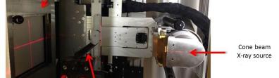
Scanner image acquisition
Scanner images acquisition is based on the Cone Beam Computed Tomography technique which requires a cone beam X-ray source, a plane detector and a computer source to process the images.
Small animal irradiation planning
The images acquired are used to plan irradiation using the Muriplan treatment planning system. Provided with the SARRP, this software allows a very fast dose planning, fusion images, organ contouring, calculates the isodoses and draws dose-volume histograms.
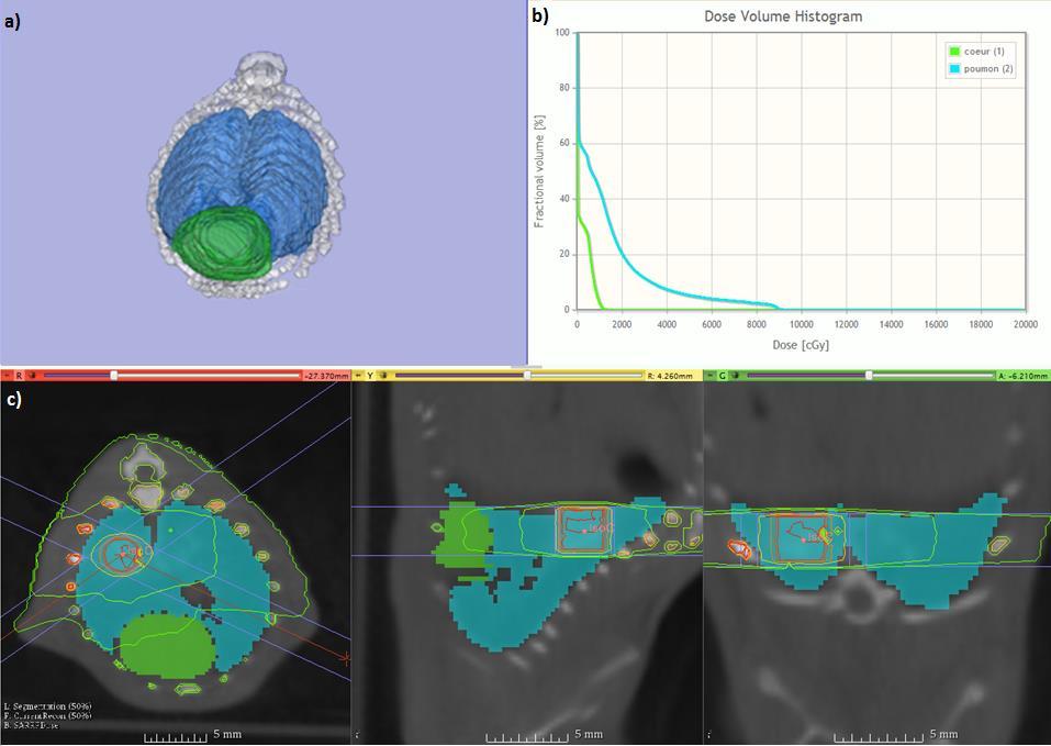
Irradiation planning consists of targeting a volume inside the animal on the scanner images and of defining the desired irradiation dose at that point. Dose distribution is calculated in the target volume and in the adjacent organs (Figure 2). Each treatment plan is unique and used to accurately target the irradiation area.
Other irradiation types
The SARRP is mainly used for highly localized irradiation on small animals. However, by removing the additional collimators, much larger irradiation fields can be used (up to 12.5 x 12 .5 cm² at the isocenter) especially for cellular irradiation.
This irradiator comes in addition to the ALPHÉE (medical accelerator, high-energy facility) and MIRCOM (ion microbeam) facilities, especially in terms of energy and type of radiation used at the IRSN.
Related research
The IRSN uses the SARRP irradiator in several types of experimental radiobiology research programs.
It especially contributes to research on the potential adverse effects of interventional radiology. This type of radiology, which consists of performing radiological image-guided and image-controlled invasive medical procedures, can have adverse effects such as radiological burns. The IRSN is seeking to more effectively characterize this type of lesion to be able to develop cell therapy treatments.
The SARRP is also used for the fine analysis of the adverse effects of high ionizing radiation doses delivered during certain types of radiotherapy. It involves studying the effects of highly localized radiation on healthy organs surrounding the tissue treated for cancer. The IRSN more specifically looks at the potential damage to the lungs (in stereotactic conditions), the bladder and bone. As for the effects of interventional radiology, one of the medium-term objectives is to better understand the mechanisms of development of radiation-induced lesions in healthy tissue and to be able to develop therapeutic strategies, especially using cell therapy.
The SARRP is also used to study the effects of low radiation doses (≤ 2 Gy). The consequences of this type of exposure on brain function and on the related pathophysiological mechanisms are studied through the development of several irradiation models on the facility (highly localized irradiation of certain cerebral sub-structures and of the whole head).
Lastly, the irradiator is used by the IRSN as part of the ROSIRIS program, the purpose of which is to better understand the causal relationship between the early effects and the delayed effects of ionizing radiation on the body. The type of initial radiation-induced cell damage depends on the way in which the energy of an ionizing particle is deposited within a cell, and therefore the physical characteristics of that particle. To analyze the damage, one of the research avenues consists of establishing links between biophysical modeling of early radiation-induced events and their later consequences on molecules and cells, by studying the distribution, signaling and repair of the initial DNA damage and restoration of the chromatin architecture according to the type of ionizing radiation.
The SARRP offers reliable and reproducible dosimetry, with currently (September 2017), around ten dosimetry protocols available for in vitro irradiation, for voltages between 40 and 220 kV and intensities of between 2.5 and 27 mA.
The experiments are conducted in accordance with the set procedures and animal testing ethics rules.
Technical specifications